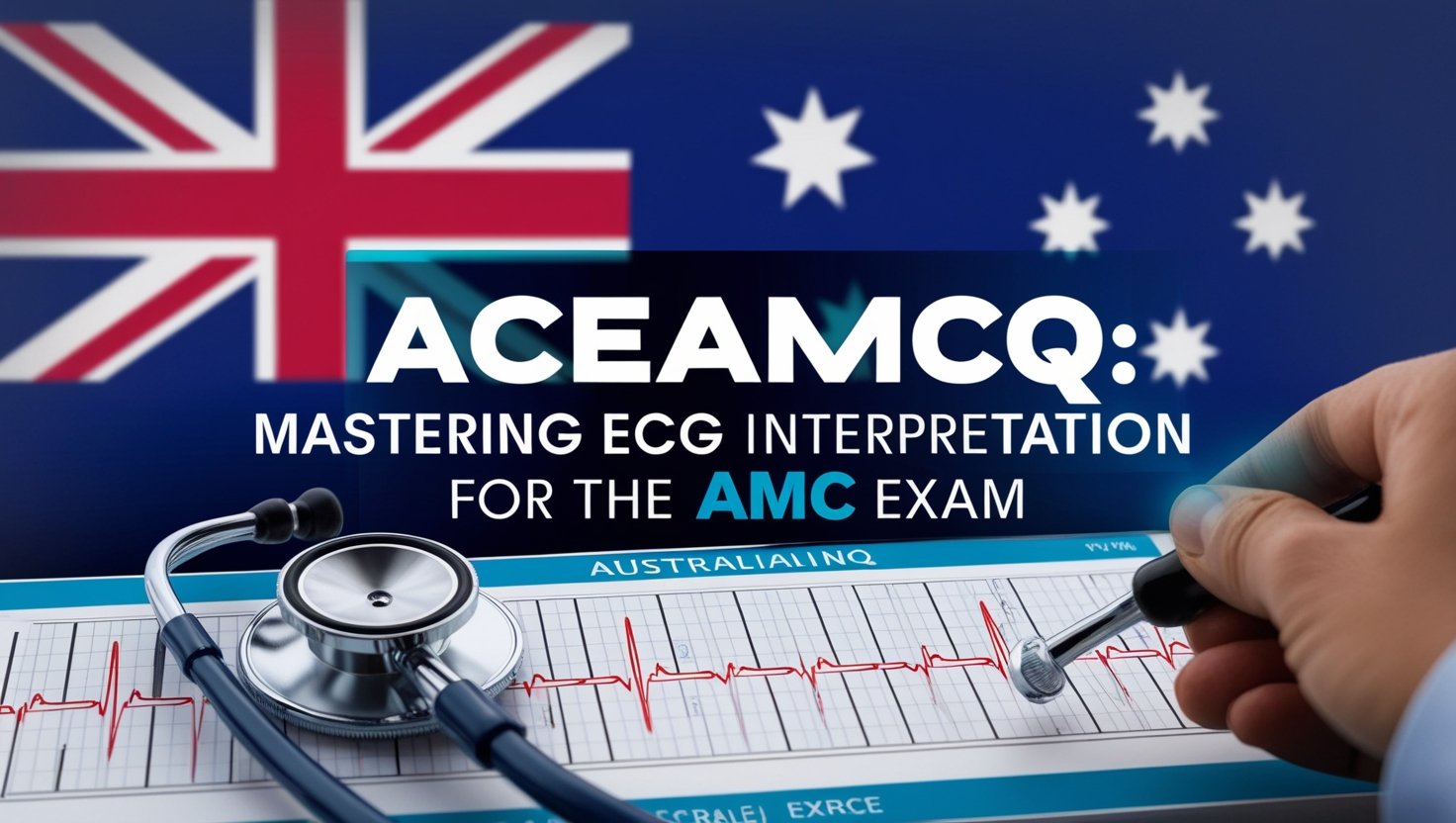
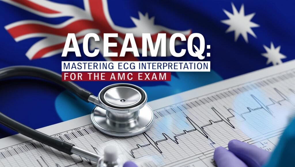
Electrocardiography (ECG) interpretation is a fundamental skill for medical professionals, especially those preparing for the Australian Medical Council (AMC) exam. Given its significance in diagnosing and managing cardiac emergencies, mastering ECG interpretation can be a game-changer in acing the AMC exam. This article will guide you through key aspects of ECG interpretation, common cardiac emergencies, and essential tips to enhance your performance in the exam.
Understanding ECG Basics
An ECG records the electrical activity of the heart and provides crucial information about heart rhythm, conduction pathways, and potential abnormalities. A standard ECG consists of 12 leads, each offering a different perspective of the heart’s electrical activity.
ECG Waves and Intervals
To interpret an ECG accurately, it is essential to understand its basic components:
- P wave: Represents atrial depolarization.
- PR interval: Indicates the time taken for the electrical impulse to travel from the atria to the ventricles (normal range: 120-200 ms).
- QRS complex: Represents ventricular depolarization (normal duration: <120 ms).
- ST segment: Indicates early ventricular repolarization; elevation or depression may suggest ischemia or infarction.
- T wave: Represents ventricular repolarization.
- QT interval: Measures the total time for ventricular depolarization and repolarization; a prolonged QT can indicate a risk of arrhythmias.
Common Cardiac Emergencies and Their ECG Findings
1. Myocardial Infarction (MI)
Myocardial infarction, or heart attack, occurs due to an obstruction in the coronary arteries, leading to ischemia and necrosis of heart tissue.
ECG Findings:
- ST-segment elevation (STEMI) in at least two contiguous leads
- ST-segment depression and T-wave inversion (NSTEMI)
- Pathological Q waves in old infarctions
2. Arrhythmias
Arrhythmias are abnormal heart rhythms that can be life-threatening.
Common Types:
- Atrial fibrillation (AF): Irregularly irregular rhythm, absent P waves, and fibrillatory waves.
- Atrial flutter: “Sawtooth” flutter waves, typically at a rate of 250–350 bpm.
- Ventricular tachycardia (VT): Broad QRS complexes with a rate >100 bpm.
- Ventricular fibrillation (VF): Chaotic electrical activity, no identifiable P waves, QRS complexes, or T waves.
3. Heart Blocks
Heart blocks result from impaired conduction in the AV node or below.
- First-degree AV block: Prolonged PR interval (>200 ms) but no missed beats.
- Second-degree AV block (Mobitz type I and II): Progressive PR prolongation with a dropped beat (Type I) or sudden dropped QRS without PR prolongation (Type II).
- Third-degree (complete) AV block: No association between P waves and QRS complexes, requiring immediate intervention.
4. Pulmonary Embolism (PE)
Pulmonary embolism can present with various ECG changes, though none are entirely specific.
ECG Findings:
- S1Q3T3 pattern (Deep S in lead I, Q wave in lead III, and T-wave inversion in lead III)
- Right bundle branch block (RBBB)
- Sinus tachycardia (most common finding)
5. Hyperkalemia and Hypokalemia
Electrolyte imbalances significantly affect ECG findings.
- Hyperkalemia: Tall peaked T waves, widened QRS, absent P waves, and sine-wave pattern in severe cases.
- Hypokalemia: Flattened T waves, U waves, and prolonged QT interval.
ECG Interpretation Strategies for the AMC Exam
1. Follow a Systematic Approach
A structured approach helps ensure no crucial findings are missed. Follow these steps:
- Check patient details and clinical context.
- Assess rate and rhythm.
- Analyze P waves and PR interval.
- Examine QRS complex duration and morphology.
- Evaluate ST segments and T waves.
- Identify any additional findings (e.g., Q waves, U waves).
2. Recognize Patterns and Clinical Correlations
Pattern recognition is crucial for ECG interpretation. Linking ECG findings with clinical presentations (e.g., chest pain with ST elevation suggests MI) improves diagnostic accuracy.
3. Practice with Real Cases and ECG Tracings
The AMC exam includes ECG-based questions, so practice interpreting a variety of ECGs. Use online resources, textbooks, and mock exams to build confidence.
4. Master Time-Sensitive Diagnoses
Some ECG findings indicate immediate life-threatening conditions, such as:
- STEMI requiring urgent reperfusion therapy
- VT/VF needing defibrillation
- Complete heart block requiring pacing
- Hyperkalemia requiring urgent correction
5. Stay Updated with Guidelines
Familiarize yourself with the latest Australian guidelines on cardiac emergencies, such as those from the National Heart Foundation of Australia.
Conclusion
ECG interpretation is a vital skill for medical professionals, particularly for those preparing for the AMC exam. A thorough understanding of ECG basics, recognition of life-threatening cardiac emergencies, and systematic interpretation strategies can significantly enhance performance. Regular practice with real ECG tracings and staying updated with guidelines will not only help in passing the AMC exam but also in delivering effective patient care in real-life clinical scenarios. Read more blog…